

 , Şeyda Dereli 2
, Günseli Miray Özdemir 2
, Utku Uluköksal 2
, Fatma Can 2
, Can Yücel Karabay 2
, Şeyda Dereli 2
, Günseli Miray Özdemir 2
, Utku Uluköksal 2
, Fatma Can 2
, Can Yücel Karabay 2
2Department of Cardiology, Dr. Siyami Ersek Thoracic and Cardiovascular Training and Research Hospital, Türkiye
Abstract
Background: Coarctation of the aorta (CoA) is a congenital obstructive anomaly that may remain undiagnosed until adulthood, often presenting with systemic hypertension. While percutaneous interventions, including balloon angioplasty and stent implantation, are increasingly utilized in adults, real-world data on procedural outcomes, complications, and long-term mortality remain limited. This study aimed to evaluate procedural outcomes, complication rates, and short- and long-term mortality associated with percutaneous interventions in adult patients with CoA.
Methods: A single-center, retrospective observational study was conducted, including adult patients with CoA who underwent percutaneous intervention between July 2017
and July 2023. Procedural success was defined as a residual peak systolic gradient ≤20 mmHg without major complications. Patient demographics, comorbidities, procedural details, complications, and follow-up outcomes were analyzed.
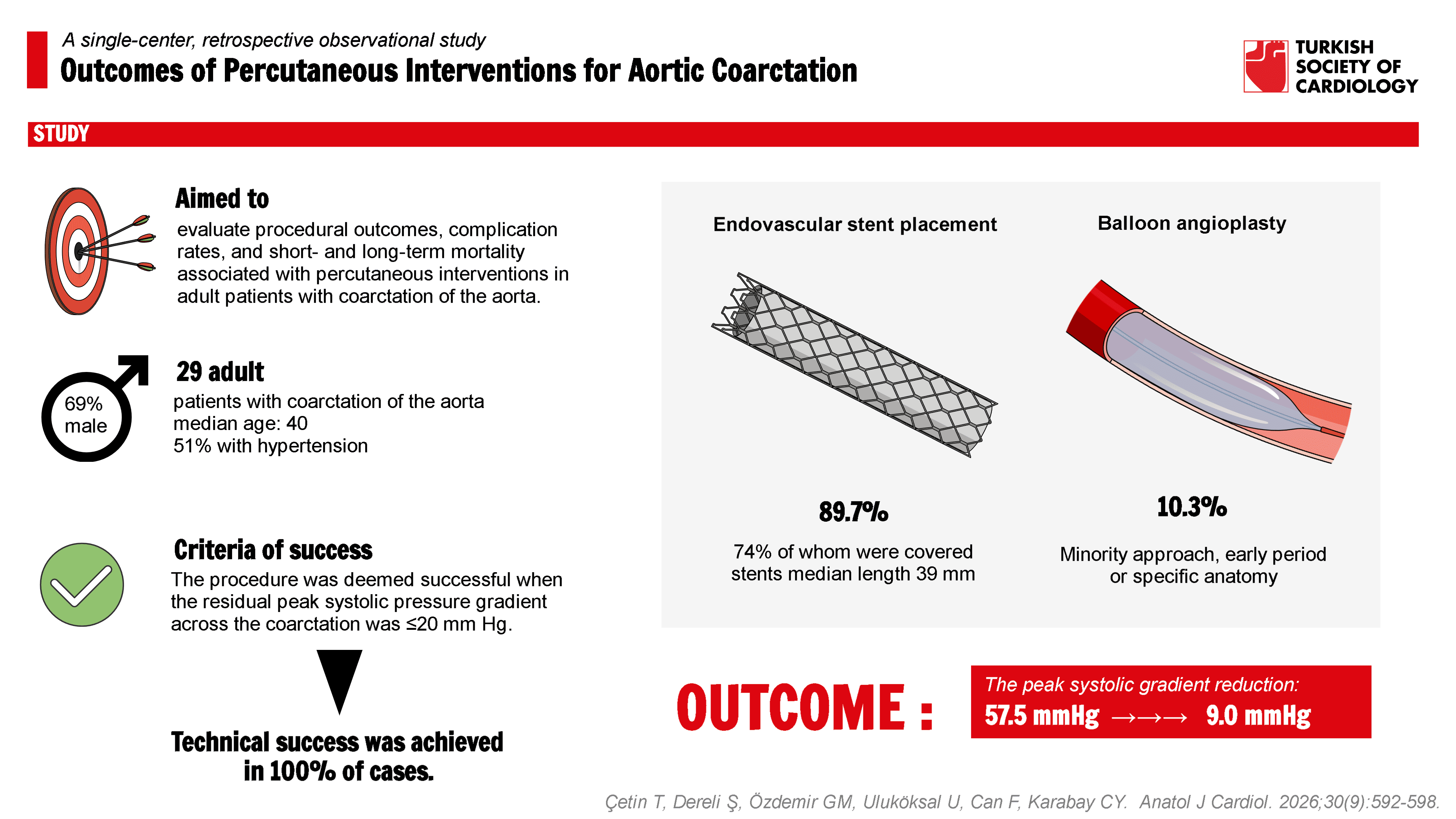
Results: Twenty-nine patients (median age 40 [29-45] years; 31% female) were included. Endovascular stent placement was performed in 89.7% of patients, 74% of whom were covered stents (mean length 39 mm). Balloon angioplasty alone was used in 10.3% of cases. The procedure was associated with a marked reduction in the peak systolic gradient, decreasing from a median of 57.5 mmHg [50.0-68.8] before the procedure to 9.0 mmHg [1.2-11.8] after the procedure. Complications occurred in 6.9%, including 1 aortic rupture and 1 access site bleeding. Recoarctation developed in 14% of patients at a median of 17.5 months. No in-hospital deaths occurred; out-of-hospital mortality was 6.9%, including 1 death of unknown cause and 1 following posterior mediastinal hematoma after aortic rupture.
Conclusions: Percutaneous interventions in adult CoA achieve favorable procedural success and low in-hospital mortality, while rare but serious complications underscore the necessity for meticulous management and lifelong follow-up.
Graphical Abstract
Highlights
- Percutaneousinterventions for adult coarctation of the aorta demonstrated high proceduralsuccess and low in-hospital complication rates.
- Endovascular stent implantation was thepredominant treatment strategy and effectively reduced post-procedural pressuregradients.
- Lifelong surveillance remains essentialbecause of the risk of late recoarctation and vascular events
Introduction
Coarctation of the aorta (CoA) is a congenital obstructive anomaly, most commonly involving a narrowing of the aortic segment distal to the left subclavian artery, with an incidence ranging between 0.06% and 0.08%.
This report assessed the impact of percutaneous interventions on procedural outcomes, complication rates, early and late mortality in cases of interrupted aortic arch or aortic coarctation in a tertiary referral hospital.
Methods
Ethical Statement
This was a retrospective observational study, and no additional interventions beyond standard clinical care were performed for research purposes. Ethical approval for the study was granted by the Ethics Committee of the University of Health Sciences (document registration number: 23/431).
Study Population and Design
A single-center retrospective observational study analysis was conducted on patients admitted to the institution with aortic coarctation who underwent percutaneous intervention over a 6-year period between July 2017 and July 2023. All procedures performed in accordance with current indications during this period were included, and follow-up assessments focused on complications, procedural success, and their impact on mortality. Patients younger than 18 years were excluded from the study. Patient demographics, comorbid conditions, laboratory results, treatment details, follow-up information after discharge, and outcome-related clinical parameters were collected retrospectively from hospital records, the institutional electronic database, and national health registries, including the Turkish Ministry of Health’s e-Nabız and Medula systems.
Hypertension was defined as a systolic blood pressure ≥140 mmHg and/or diastolic blood pressure ≥90 mm Hg, measured at the brachial artery after participants had rested for at least 5 minutes in a seated position.
A diagnosis of CoA was confirmed in individuals with systemic hypertension accompanied by a systolic pressure difference of at least 20 mmHg between the upper and lower extremities, as validated by imaging modalities such as aortic computed tomography angiography (CTA), aortography, or echocardiography. All patients considered for inclusion underwent pre-procedural aortic CTA to ensure accurate diagnosis of CoA, detailed assessment of the aortic anatomy, and appropriate determination of stent and balloon dimensions.
Procedure
All interventions were carried out by 2 experienced operators under deep sedation with anesthetic support in the cardiac catheterization laboratory. Upon achieving femoral access, anticoagulation was initiated with heparin at a dose of 100 IU/kg, or 5000 IU in adult patients. In all participants, fluoroscopy guided the puncture of the common femoral artery, allowing placement of a 6-F introducer sheath. A right radial artery approach was additionally utilized to enhance visualization of the coarctation region and surrounding anatomy, facilitating accurate positioning of the device. A 0.035-inch hydrophilic guidewire was advanced across the coarctation with catheter support, typically using either a multipurpose or Judkins right catheter (Cordis Corporation, Florida). This was subsequently exchanged for a 0.035-inch Amplatz super-stiff guidewire (Cook Cardiology, Indiana) and the introducer sheath was upsized to 12-F. Prior to stent implantation, the peak systolic gradient was assessed using invasive hemodynamic measurements. For stent deployment, a balloon-in-balloon technique was used, which allowed controlled and precise expansion for both covered and non-covered stents. Stent diameter selection was based on the diameter of the descending thoracic aorta at the diaphragmatic level, which was used as the anatomical reference. Precise stent positioning was confirmed with aortography obtained in several projections to optimally visualize the lesion. Rapid ventricular pacing was not used during stent implantation in the study cohort. Following control angiography to assess stent position, expansion and potential vascular complications, the procedure was concluded once proper stent placement and adequate reduction of the pressure gradient were confirmed.
The procedure was deemed successful when the residual peak systolic pressure gradient across the coarctation was ≤20 mm Hg, and no complications such as stent migration or aortic dissection were observed.
Laboratory, Echocardiographic, and Aortic Computed Tomography Angiography Assessments
Laboratory evaluation included complete blood count, liver and kidney function tests, serum electrolytes, lipid profile, plasma glucose, and thyroid function parameters. Blood counts were determined using an automated hematology analyzer (MINDRAY BC-6800, China), and biochemical assays were carried out with the ARCHITECT PLUS CI-4100 platform (Abbott, USA).
All participants underwent transthoracic echocardiography (TTE) performed by an experienced cardiologist. Examinations were performed both before the procedure and at follow-up evaluations. This assessment also enabled the detection of concomitant intracardiac abnormalities. Follow-up echocardiographic evaluations, performed after the intervention and during subsequent clinical visits, were utilized to verify stent position and to evaluate for residual gradients. The echocardiographic protocol included measurements of left ventricular ejection fraction (LVEF), left ventricular end-diastolic diameter (LVEDD) and left ventricular end-systolic diameter (LVESD), interventricular septal thickness, and posterior wall thickness. In addition, valvular and intracardiac abnormalities were systematically assessed and documented.
Patients were followed according to guideline recommendations.
Statistical Analysis
Continuous variables are presented as mean ± standard deviation or median [Q1-Q3] (interquartile range), as appropriate. Categorical variables are expressed as counts and percentages. Normality of continuous variables was assessed using the Shapiro–Wilk test. For comparisons between 2 groups, the independent-samples
Additional exploratory analyses were performed according to sex and predefined age groups (<29, 29-45, and >45 years). Continuous variables in subgroup analyses were compared using distribution-appropriate tests, and categorical variables were compared using Fisher’s exact test. For the comparison of recoarctation rates between covered and uncovered stents, a 2-sided Fisher’s exact test was used, and effect size was reported as risk difference (RD) along with 95% CIs.
Follow-up TTE for assessment of recoarctation was available in 27/29 patients; aside from these 2 missing follow-up echocardiograms, there were no other missing follow-up data relevant to the analyses. All analyses were performed using R (R Foundation for Statistical Computing, Vienna, Austria). A 2-sided
Results
Between July 2017 and July 2023, 29 patients with CoA were treated at the center. The median age was 40 [29-45] years, with more than half of the patients between 29 and 45 years, and 31% were female. In the present study, endovascular stent placement was the predominant interventional approach for CoA, performed in 89.7% of cases, whereas balloon angioplasty alone was utilized in a minority of patients, 10.3%.
In sex-based comparisons, male patients had a higher pre-procedural peak gradient than female patients (64.2 ± 16.9 vs. 49.9 ± 10.8 mmHg,
In exploratory analyses according to predefined age groups (<29, 29-45, and >45 years), coronary artery disease and distal aortic diameter differed significantly across groups. Coronary artery disease was present only in patients aged >45 years (33.3%,
Procedure-related complications occurred in 6.9% of patients, including 1 major complication consisting of aortic rupture (3.4%) and 1 minor complication consisting of bleeding at the closure device access site (3.4%). At a median follow-up of 6 months (IQR 4.7-10.7) echocardiography was available for all 17 patients. The median LVEDD was 45.5 [42.0-52.5] mm, LVESD 30.0 [27.0-34.0] mm, and LVEF 60% [60-60]. Doppler-derived peak gradient across the treated segment was 9.0 [1.2-11.8] mmHg. Re-coarctation occurred in 4 patients (14%), with a median time to diagnosis of 17.5 months (IQR 11.0-47.3), and was confirmed in 2 of 20 patients in the covered stent group and in 2 patients in the uncovered group; this difference was not statistically significant (Fisher’s exact test,
Discussion
This single-center retrospective observational study evaluated the procedural outcomes, complication rates, and long-term follow-up results of percutaneous interventions for adult patients with CoA. Advances in technology and treatment options have led to increased life expectancy in patients with congenital heart disease. Individuals living with aortic coarctation are increasingly being diagnosed at older ages. In the study, the mean age of patients was higher compared to that reported in previous studies.
These favorable outcomes can be attributed in part to the widespread adoption of endovascular stenting as the primary treatment strategy, while balloon angioplasty was reserved for selected cases. Covered stents were employed in the majority of patients, reflecting contemporary guideline recommendations that favor their use in adults due to their potential to reduce recoarctation and vascular injury. Balloon angioplasty as a standalone procedure often results in residual or recurrent obstruction.
Sadeghipour et al
Early and late complications remain an important clinical concern. Two procedure-related complications were observed—1 aortic rupture and 1 access site bleeding. The patient who experienced aortic rupture subsequently developed a posterior mediastinal hematoma and died within the first month after the intervention. This rupture did not occur during the procedure or index hospitalization but developed approximately 1 month after discharge in a patient treated with a covered stent, in whom no predilatation or intraprocedural complications were observed. This delayed presentation suggests that the underlying mechanism was unlikely to be related to acute procedural factors such as balloon-to-aorta diameter mismatch or excessive overdilatation. Instead, delayed aortic rupture may reflect intrinsic aortic wall fragility, chronic degenerative changes at the coarctation site, or progressive mechanical stress at the stent edges following restoration of normal aortic pressures. Abrupt normalization of afterload and altered wall shear stress after relief of severe coarctation may also contribute to delayed weakening of the aortic wall. Although covered stents are designed to reduce the risk of acute rupture, they may not fully prevent late vascular complications in patients with advanced aortic wall pathology. A second death occurred about 3 months after the intervention, though the available data did not clarify the cause. In the study by Moltzer et al,24 acute complications included 1 death from aortic rupture and 2 groin hematomas, while late complications involved stent migration to the ascending aorta, pseudoaneurysm formation at the initial stent site, and external iliac artery occlusion. In the study, recoarctation occurred in approximately 14% of patients during follow-up, with a median time to diagnosis of 18 months. This incidence aligns with prior studies; however, when considering studies that focused exclusively on adult patients, such as that by Sadeghipour et al,21 recoarctation occurred in fewer than 10% of patients. Recoarctation may arise from stent underexpansion, neointimal proliferation, or vascular remodeling, particularly in cases with complex anatomy. According to Zussman et al,
Study Limitations
This study is limited by its retrospective, single-center design, which may introduce selection and information biases and limit generalizability. The relatively small sample size further restricts statistical power and the applicability of findings to broader populations. In addition, follow-up duration was variable and limited, potentially underestimating late complications or re-coarctation events. Follow-up was primarily based on TTE and office blood pressure measurements, without routine use of advanced imaging or ambulatory blood pressure monitoring, which may have limited the detection of late vascular complications and persistent hypertension. In the future, larger prospective multicenter studies with long-term follow-up could provide more robust insights into the durability and outcomes of aortic coarctation interventions.
Conclusion
In the present study, percutaneous interventions for adult CoA were associated with a high rate of procedural success and low in-hospital mortality. Endovascular stent implantation was the predominant therapeutic strategy and was associated with a reduction in post-procedural pressure gradients. Although complications were uncommon, serious adverse events such as aortic rupture underscore the necessity of meticulous procedural planning and execution. Furthermore, while rare, the occurrence of recoarctation highlights the importance of lifelong cardiologic surveillance to ensure timely detection and management of recurrent aortic obstruction. Overall, these findings support percutaneous endovascular therapy as a viable treatment modality for adult patients with coarctation of the aorta, emphasizing the critical role of technical precision and continuous follow-up in optimizing long-term outcomes.
No artificial intelligence (AI) or AI-assisted technologies were used in the preparation of this study.
Footnotes
References
- Amato JJ, Douglas WI, James T, Desai U. Coarctation of the aorta. Semin Thorac Cardiovasc Surg Pediatr Card Surg Annu. 2000;3():125-141.
- Law MA, Collier SA, Sharma S, Tivakaran VS. Coarctation of the aorta. 2024 Dec 11. . ;():-.
- Brown ML, Burkhart HM, Connolly HM. Coarctation of the aorta: lifelong surveillance is mandatory following surgical repair. J Am Coll Cardiol. 2013;62(11):1020-1025.
- Kenny D, Hijazi ZM. Coarctation of the aorta:from fetal life to adulthood. Cardiol J. 2011;18(5):487-495.
- Kische S, D'Ancona G, Stoeckicht Y, Ortak J, Elsässer A, Ince H. Percutaneous treatment of adult isthmic aortic coarctation: acute and long-term clinical and imaging outcome with a self-expandable uncovered nitinol stent. Circ Cardiovasc Interv. 2015;8(1):-.
- Jurcut R, Daraban AM, Lorber A. Coarctation of the aorta in adults: what is the best treatment? Case report and literature review. J Med Life. 2011;4(2):189-195.
- Sohrabi B, Jamshidi P, Yaghoubi A. Comparison between covered and bare Cheatham-Platinum stents for endovascular treatment of patients with native post-ductal aortic coarctation: immediate and intermediate-term results. JACC Cardiovasc Interv. 2014;7(4):416-423.
- Meadows J, Minahan M, McElhinney DB, McEnaney K, Ringel R. Intermediate outcomes in the prospective, multicenter coarctation of the aorta stent trial (COAST). Circulation. 2015;131(19):1656-1664.
- Sadeghipour P, Mohebbi B, Firouzi A. Balloon-expandable Cheatham-platinum stents versus self-expandable nitinol stents in coarctation of aorta: A randomized controlled trial. JACC Cardiovasc Interv. 2022;15(3):308-317.
- McEvoy JW, McCarthy CP, Bruno RM. 2024 ESC Guidelines for the management of elevated blood pressure and hypertension. Eur Heart J. 2024;45(38):3912-4018.
- Mazzolai L, Teixido-Tura G, Lanzi S. 2024 ESC Guidelines for the management of peripheral arterial and aortic diseases. Eur Heart J. 2024;45(36):3538-3700.
- Mohammadzadeh Shabestari M, Eshraghi A, Hakim Attar F. Evaluation of short and mid-term clinical outcomes in patients with aortic coarctation treated with self-expandable stents. Sci Rep. 2024;14(1):-.
- Yazıcı HU, Göktekin O, Ulus T. Erişkinlerde aort daralmasının stent ile tedavisine ilişkin ilk deneyimlerimiz. Turk Kardiyol Dern Ars. 2011;39(3):214-218.
- Erdem A, Akdeniz C, Sarıtaş T. Cheatham-Platinum stent for native and recurrent aortic coarctation in children and adults: immediate and early follow-up results. Anadolu Kardiyol Derg. 2011;11(5):441-449.
- Rao PS, Thapar MK, Kutayli F, Carey P. Causes of recoarctation after balloon angioplasty of unoperated aortic coarctation. J Am Coll Cardiol. 1989;13(1):109-115.
- Pourmoghadam KK, Velamoor G, Kneebone JM, Patterson K, Jones TK, Lupinetti FM. Changes in protein distribution of the aortic wall following balloon aortoplasty for coarctation. Am J Cardiol. 2002;89(1):91-93.
- Fawzy ME, Sivanandam V, Galal O. One- to ten-year follow-up results of balloon angioplasty of native coarctation of the aorta in adolescents and adults. J Am Coll Cardiol. 1997;30(6):1542-1546.
- Koerselman J, de Vries H, Jaarsma W, Muyldermans L, Ernst JM, Plokker HW. Balloon angioplasty of coarctation of the aorta: a safe alternative for surgery in adults: immediate and mid-term results. Catheter Cardiovasc Interv. 2000;50(1):28-33.
- Rodés-Cabau J, Miró J, Dancea A. Comparison of surgical and transcatheter treatment for native coarctation of the aorta in patients > or = 1 year old. Am Heart J. 2007;154(1):186-192.
- Taggart NW, Minahan M, Cabalka AK. Immediate outcomes of covered stent placement for treatment or prevention of aortic wall injury associated with coarctation of the aorta (COAST II). JACC Cardiovasc Interv. 2016;9(5):484-493.
- Sadeghipour P, Pouraliakbar HR, Farrashi M. Balloon-expandable versus self-expanding stents in native coarctation of the aorta: three-year results of a randomised controlled trial. EuroIntervention. 2024;20(9):613-615.
- Musto C, Cifarelli A, Pucci E. Endovascular treatment of aortic coarctation: long-term effects on hypertension. Int J Cardiol. 2008;130(3):420-425.
- Boshoff D, Budts W, Mertens L. Stenting of hypoplastic aortic segments with mild pressure gradients and arterial hypertension. Heart. 2006;92(11):1661-1666.
- Moltzer E, Roos-Hesselink JW, Yap SC. Endovascular stenting for aortic (re)coarctation in adults. Neth Heart J. 2010;18(9):430-436.
- Zussman ME, Hirsch R, Herbert C, Stapleton GE. Transcatheter intervention for coarctation of the aorta. Cardiol Young. 2016;26(8):1563-1567.